Approach
Electrophysiology
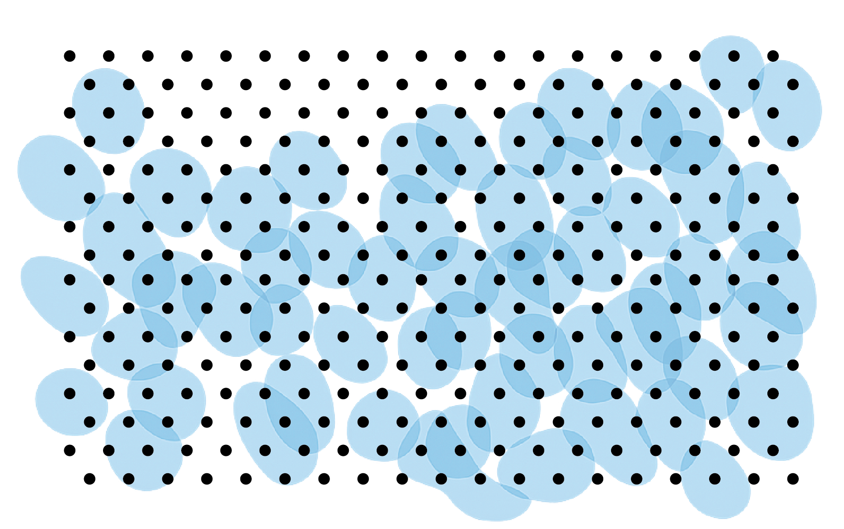
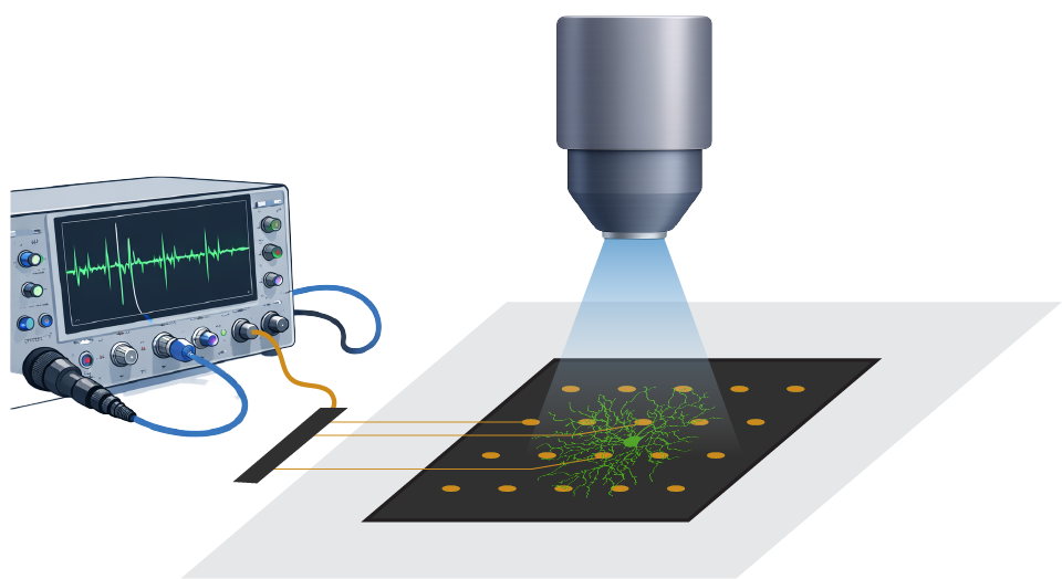
We use large-scale electrophysiology to record from thousands of retinal ganglion cells at once. These recordings capture how different types of ganglion cells encode and transmit visual information to the brain, in normal and diseased states.
Viral vectors
We work with institutional cores to design custom viral constructs for our projects. These vectors enable us to target cell types in the retina and in the brain for anatomical tracing, circuit mapping, and functional characterization.

Optogenetics
We use optogenetic tools to manipulate and measure the activity of cell types. These tools are developed and validated in murine and large-animal models for both fundamental retinal research and translational studies.
Light-sheet imaging
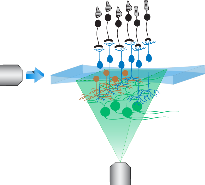
Built on principles of restricted planar imaging, light-sheet fluorescence microscopy enables measurements of neural activity at single-cell resolution across large regions of the ex vivo retina (read more). For functional characterization, we target retinal interneurons and ganglion cells for GCaMP expression. We are also refining these tools to study signal propagation within circuits, and within neuronal compartments such as dendrites and axons.
Molecular assays
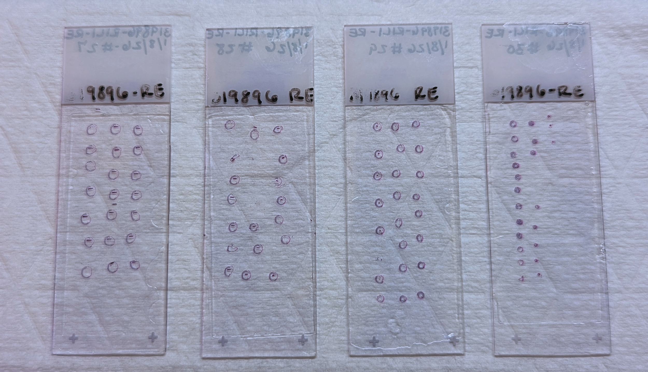
We use various molecular assays such as qPCR, immunofluorescence, immunohistochemistry, and targeted transcript panels to measure changes in gene and protein expression in retinal and brain tissues.
Transcriptomics
To map gene expression during retinal development or during retinal degeneration, we take advantage of high-throughput sequencing methods. This generates high-resolution molecular atlases of specific cell types and their circuit interactions. We expect the data to offer insights into the genes that are regulated in blinding diseases and how they can be targeted for therapies.